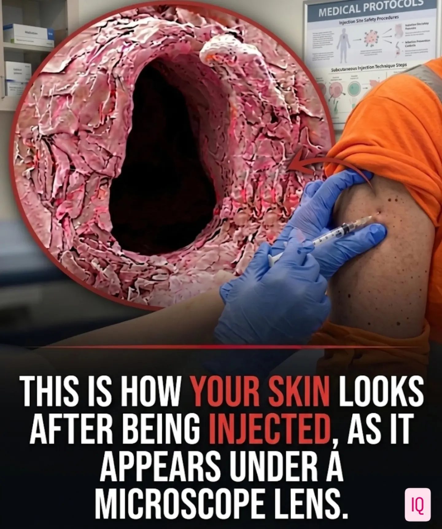
Skin Injection Visualization
The image of skin under a microscope after being injected is fascinating, revealing the intricacies of our body's structure. This visualization, often misinterpreted, provides valuable insights into how skin responds to minor punctures.
Context / Why This Matters
Understanding how skin tissue reacts to injections is crucial for medical professionals and researchers. This knowledge aids in developing more effective and less invasive medical procedures, as well as advancements in tattooing and other dermal applications. By examining the microscopic details of skin after injection, we can better understand the body's healing process and improve treatment methods.
Main Discussion
The Misconception
Social media often misrepresents images of skin under a microscope. Many posts falsely claim that these images depict the aftermath of a standard medical syringe injection. However, the reality is quite different. The image in question is a false-color scanning electron micrograph (SEM), captured by Anne Weston at the Francis Crick Institute. This image shows a microscopic puncture wound in the epidermis caused by a tattoo needle, magnified approximately 280 times. The confusion arises from the dramatic visualization, which appears to show a gaping wound, but it is actually a highly magnified view of a small, localized injury.
Anatomy of the Skin
The stratum corneum, the outermost layer of the skin, is composed of flat, keratin-packed cells called corneocytes. These cells are constantly shedding, a process that helps maintain the skin's barrier function. The rough, layered texturing surrounding the cavity in the image represents this stratum corneum, providing a clear view of the skin's protective layer and its response to injury.
Scanning Electron Microscopy
Scanning electron microscopy (SEM) is a powerful tool for visualizing biological samples at high magnification. To prepare a sample for SEM, it must be completely dehydrated, preserved, and structurally stabilized in a vacuum chamber. This process gives the tissue a distinctly rigid and dry appearance, which is quite different from how a living, fluid-hydrated human puncture would look in real-time. The false-color SEM image enhances the contrast and detail, making it easier to study the microscopic structure of the skin.
The Internal Changes After Injection
When a substance is injected into the skin, the internal changes are both immediate and complex. The magnification in the visualization provides a detailed view of how the skin's layers respond to the injection. The syringe's needle creates a small puncture, and the substance injected causes local tissue deformation. The visualization shows how the stratum corneum and underlying layers react, providing valuable insights into the body's response to injections.
Practical Tips
Understanding Visualizations
When viewing medical visualizations, it's essential to understand the context and the methods used to create the image. Always consider the magnification level, the preparation techniques used, and the specific type of microscopy employed. This knowledge helps in interpreting the image accurately and avoiding misconceptions.
Importance of Correct Information
Misinterpretations of medical images can lead to misunderstandings and unnecessary fears. It's crucial to verify the source and the context of any medical visualization. Always look for trusted sources and consider the expertise of the person or institution providing the information.
Application in Medical Practice
For medical professionals, understanding the microscopic structure of the skin is vital for accurate diagnosis and treatment. This knowledge helps in developing more precise and less invasive medical procedures, ensuring better patient outcomes.
Important Takeaways
- Misconceptions: Social media often misrepresents medical visualizations. The image in question shows a tattoo needle puncture, not a syringe injection.
- Skin Anatomy: The stratum corneum is the outermost layer of dead skin cells, providing a protective barrier and constantly renewing.
- Scanning Electron Microscopy: This technique involves dehydration and vacuum stabilization, giving the tissue a rigid and dry appearance.
- Internal Changes: The visualization shows how the skin's layers react to an injection, providing valuable insights into the body's healing process.
Conclusion
The visualization of skin tissue under a microscope after being injected reveals the intricate details of our skin's response to punctures and injections. By understanding the context and the methods used to create these images, we can better interpret them and apply this knowledge to medical practice. Whether in research, medical procedures, or dermal applications, a clear understanding of skin tissue's microscopic structure is invaluable. This knowledge helps in developing more effective and less invasive treatments, ensuring better patient outcomes and advancements in the field.
Watch the Reel
Questions readers ask
What is the stratum corneum and why is it important in understanding skin injections?
The stratum corneum is the outermost layer of the skin, serving as a protective barrier. Observing its response to injections under a microscope helps professionals understand how the skin begins to heal from minor punctures, aiding in the development of less invasive procedures.
How does a microscopic view aid in understanding skin injections?
A microscopic view of skin tissue after an injection offers a detailed look at the body's initial response to a puncture. This visualization helps professionals comprehend the skin's self-repair process, which is crucial for advancements in medical procedures and tattooing.
What can medical professionals learn from examining skin tissue under a microscope?
By examining skin tissue under a microscope, medical professionals can gain detailed insights into the body's healing process. This includes observing how the skin's protective barrier, the stratum corneum, reacts to punctures, aiding in the creation of more effective and less invasive treatment methods.
Why is it important to study the microscopic wound healing process of the skin?
Studying the microscopic wound healing process of the skin allows professionals to understand how the skin repairs itself from minor injuries. This knowledge is beneficial for improving treatment methods, developing new procedures, and enhancing techniques in tattooing and other dermal applications.
How does a scanning electron microscope help in understanding skin injections?
A scanning electron microscope provides a high-resolution, magnified view of the skin tissue, enabling professionals to observe the intricate details of the skin's response to injections. This detailed inspection aids in understanding the healing process and developing better procedures.
What are some common misconceptions about skin injection images on social media?
Social media often misinterprets images of skin under a microscope, leading to misunderstandings about the injection process and the skin's healing abilities. Professional insights gained from accurate microscopic views are necessary to correct these misconceptions.
What are the benefits of understanding the injection process in skin tissue?
Understanding the injection process in skin tissue helps professionals develop more effective medical procedures, reduce invasiveness, and improve outcomes. This knowledge is also beneficial for advancements in tattooing and other dermal practices, ensuring safer and more precise techniques.
Related deep dives
Similar reads based on topic and creator.



Recent articles
Fresh deep dives from the latest Reels we unpacked.




Comments
Be the first to comment.