The Intricate World of Finger Anatomy
Fingers, with their complex structure, enable crucial movements, sensations, and protection. Understanding finger anatomy is essential for diagnosing hand injuries, guiding surgical procedures, and aiding in physiotherapy. This guide explores the layers of the finger from the deep skeleton to the superficial nail, highlighting the key structures and their functions.
Why This Matters
The layers of the finger work together to provide movement, sensation, protection, and blood supply. Each layer has a unique role that contributes to the overall function and health of the hand. Whether you're a healthcare professional, a student, or someone interested in the human body, understanding finger anatomy can provide valuable insights and practical applications.
Main Discussion
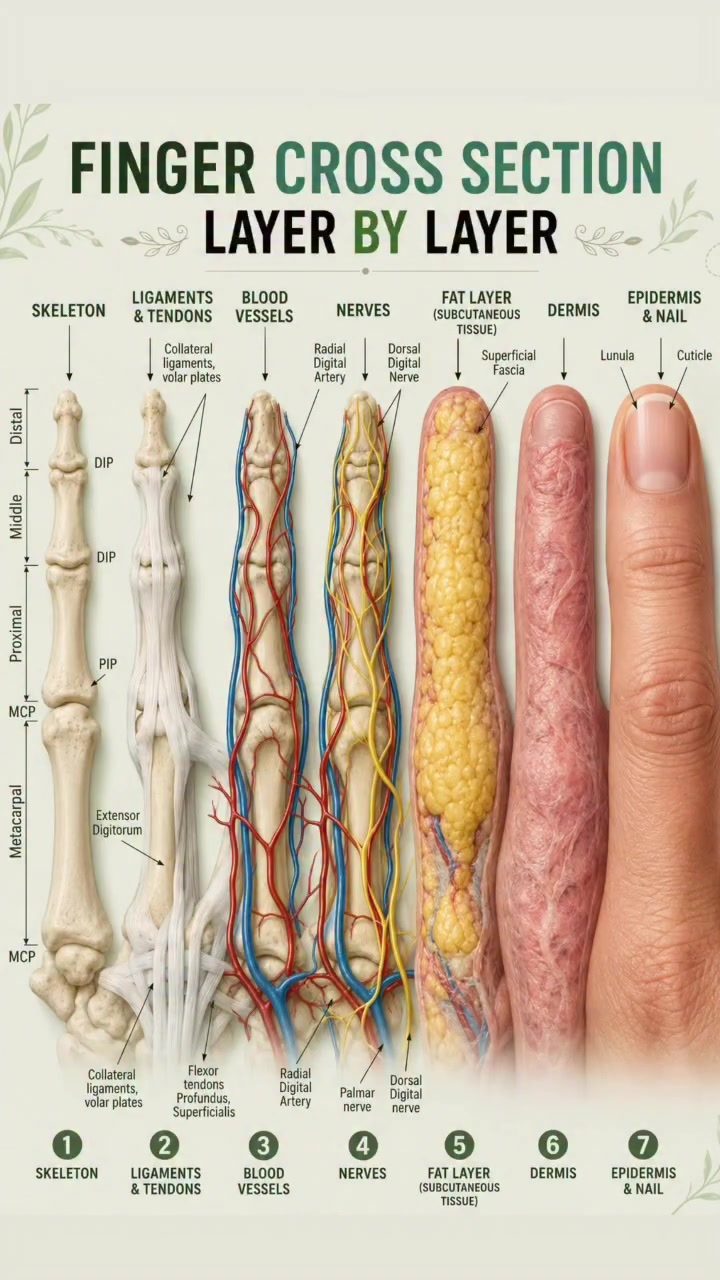
The Skeleton: Foundation and Support
The skeleton of the finger consists of phalanges and joints, which provide the structural framework for the hand. The phalanges are the bones that make up the finger, while the joints allow for movement. The metacarpal bones connect the fingers to the hand, and the distal interphalangeal (DIP) and metacarpophalangeal (MCP) joints enable bending and straightening.
Ligaments and Tendons: Stability and Movement
Ligaments and tendons play a crucial role in stabilizing the joints and enabling finger movement. Ligaments connect bone to bone, providing stability and preventing excessive movement. Tendons, on the other hand, connect muscles to bones, allowing for the movement of the fingers. The collateral ligaments and volar plates are essential for maintaining joint stability, while the extensor digitorum and flexor tendons facilitate bending and straightening of the fingers.
Blood Vessels: Oxygen and Nutrient Delivery
Blood vessels are responsible for delivering oxygen and nutrients to the finger tissues. The radial digital artery and digital nerves supply blood to the finger, while the superficial and deep veins carry blood away from the tissues. These vessels ensure that the finger remains healthy and functional.
Nerves: Sensation and Motor Control
Nerves provide sensation and control fine motor function in the fingers. The palmar digital nerves and dorsal digital nerves carry sensory information from the fingers to the brain, allowing for touch, temperature, and pain perception. These nerves also control the muscles that move the fingers, enabling precise and coordinated movements.
Fat Layer: Cushioning and Protection
The fat layer, also known as subcutaneous tissue, cushions and protects the underlying structures of the finger. This layer provides insulation and helps maintain the finger's shape and contour. It also acts as a shock absorber, protecting the bones, ligaments, and tendons from injury.
Dermis: Strength and Structural Support
The dermis is a thick layer of connective tissue that contains collagen, sweat glands, blood vessels, and sensory receptors. It provides strength and structural support to the finger, helping to maintain its shape and integrity. The dermis also plays a crucial role in wound healing and tissue repair.
Epidermis and Nail: Protection and Sensation
The epidermis is the outer layer of the finger, providing protection against injury and infection. It contains the nail, which helps to protect the fingertip and enhance sensory perception. The lunula and cuticle are important structures of the nail, contributing to its overall health and appearance.
Practical Tips
- Regular Exercise: Keeping the hands and fingers active through regular exercise can help maintain flexibility and strength.
- Proper Posture: Maintaining proper posture while using the hands can prevent strain and injury.
- Avoid Repetitive Movements: Repetitive movements can lead to tendon and nerve damage, so it's important to take breaks and vary your activities.
- Protection: Wear gloves and other protective gear when engaging in activities that could cause hand injuries.
- Proper Nutrition: Eating a balanced diet rich in vitamins and minerals can support overall hand health.
Important Takeaways
- Complex Structure: Fingers are composed of multiple specialized layers, each with unique functions.
- Critical Functions: These layers work together to provide movement, sensation, protection, and blood supply.
- Clinical Relevance: Understanding finger anatomy is crucial for diagnosing injuries, guiding surgical procedures, and supporting rehabilitation.
- Preventive Measures: Regular exercise, proper posture, and protective gear can help maintain hand health and prevent injuries.
Conclusion
Finger anatomy is a fascinating and intricate field that offers valuable insights into the human body. By understanding the layers of the finger and their functions, you can gain a deeper appreciation for the complexity and importance of hand anatomy. This knowledge can be applied in various fields, from healthcare to sports, to improve hand function and prevent injuries.
Watch the Reel
Questions readers ask
What are the main components of the finger's skeletal structure?
The finger's skeletal structure is composed of bones called phalanges, which are divided into proximal, middle, and distal phalanges. The metacarpal bones connect the fingers to the hand, providing a stable base for movement. Together, these bones form the framework that supports the hand and facilitates gripping and manipulating objects.
How do ligaments and tendons contribute to finger function?
Ligaments in the fingers connect bones to other bones, providing stability and preventing excessive movement. Tendons, on the other hand, connect muscles to bones and are crucial for finger movement. The extensor tendons straighten the fingers, while the flexor tendons bend them, enabling a wide range of motions essential for daily activities.
What is the role of blood vessels in finger anatomy?
Blood vessels in the fingers are vital for supplying oxygen and nutrients to the tissues. The digital arteries run along the sides of the fingers, branching out to form a network that ensures adequate blood flow. Proper functioning of these vessels is essential for maintaining finger health and facilitating healing in case of injuries.
Can you explain the function of the dermis and epidermis in the fingers?
The epidermis is the outermost layer of the skin, providing a protective barrier against external elements. The dermis, located beneath the epidermis, contains nerve endings, blood vessels, and sweat glands. This layer plays a crucial role in sensation, temperature regulation, and overall finger function and protection.
Why is understanding finger nerves important in hand injuries?
Nerves in the fingers, such as the digital nerves, are responsible for transmitting sensory information to the brain. Understanding the nerve structure is vital for diagnosing and treating hand injuries, as nerve damage can result in loss of sensation or motor function. This knowledge is also crucial for surgical procedures and physiotherapy to restore proper function.
What is the significance of the subcutaneous tissue in the fingers?
Subcutaneous tissue, or subcutis, is the layer of fatty tissue beneath the dermis. It serves as a cushion, protecting the deeper structures of the finger from injury. This tissue also contains blood vessels and nerves that supply the skin, playing a role in maintaining the finger's overall health and function.
How does a cross-section of a finger reveal its complex structure?
A cross-section of a finger exposes the various layers, including the nail, epidermis, dermis, subcutaneous tissue, tendons, ligaments, blood vessels, and bones. This view highlights the intricate arrangement of these components, showing how they work together to provide movement, sensation, and protection. It is a useful tool for visualizing the complexity of finger anatomy.
Related deep dives
Similar reads based on topic and creator.




Recent articles
Fresh deep dives from the latest Reels we unpacked.




Comments
Be the first to comment.