Embryo Development Under the Microscope
Embryo development is a fascinating process that starts with a single cell and progresses through various stages, eventually leading to the formation of a complex organism. Microscopic observation plays a crucial role in understanding this intricate process. The development of an embryo is a series of precise cellular events that occur in a fluid-filled environment. A microscope is an invaluable tool for scientists to observe these events in detail.
Why Microscopic Observation Matters
Microscopic observation is essential for studying embryo development because it allows researchers to examine the cellular structure and dynamics at a granular level. This detailed view helps in understanding the molecular and genetic processes that drive development. By observing the embryo under a microscope, scientists can track changes over time, identify critical developmental milestones, and intervene when necessary to study the effects of various factors.
The Process of Embryo Development
Initial Cell Injection
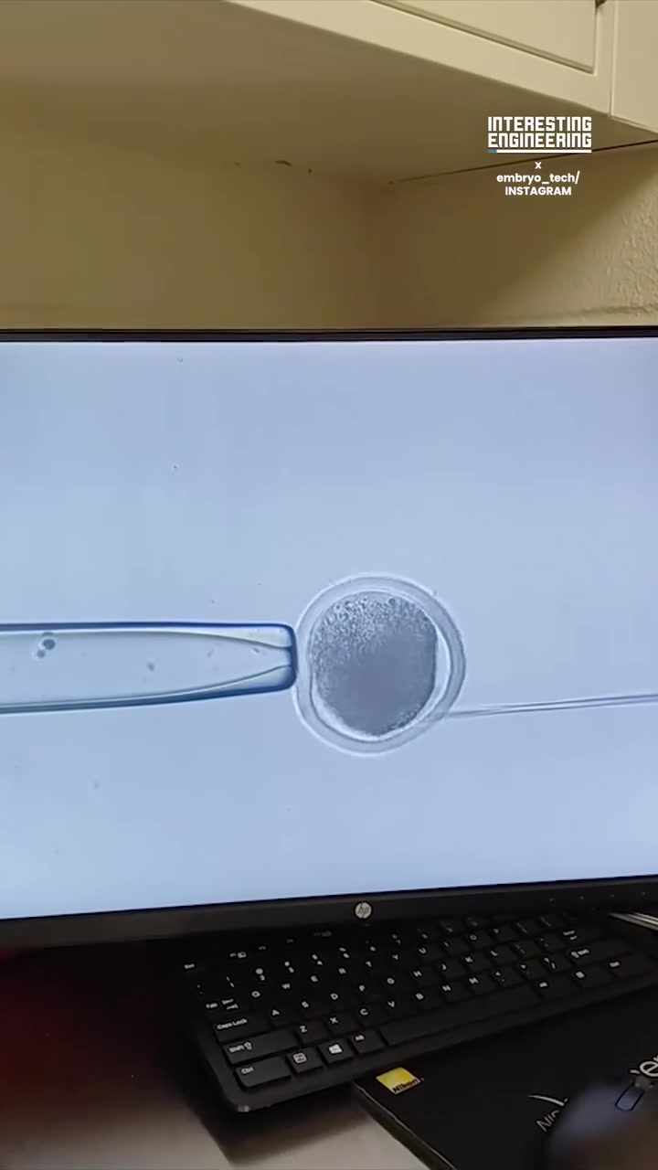
The process begins with the injection of material into a single cell using a fine needle. This step is performed with high precision to ensure that the material is deposited accurately. The injection process is crucial for introducing genetic material or other substances that can influence the embryo's development. It sets the stage for the subsequent stages of cell division and differentiation.
Time-Lapse Observation
Time-lapse imaging is a powerful technique for observing embryo development. It involves capturing a series of images at regular intervals and compiling them into a video that shows the progression of events over time. This method allows researchers to observe the dynamic process of cell division, differentiation, and movement, which are essential for the embryo's growth and development. The time-lapse images provide a detailed view of the embryo's cellular structure, helping scientists understand the complex interactions that occur during development.
Cellular Dynamics
Under a microscope, the embryo appears as a cluster of cells in a fluid-filled environment. The cells undergo rapid division and differentiation, forming distinct structures and tissues. Observing these cellular dynamics is crucial for understanding the mechanisms that govern development. Researchers can study the molecular and genetic factors that regulate cell behavior, identify key developmental pathways, and explore the effects of environmental factors on embryo development.
Practical Tips for Microscopic Observation
High-Precision Tools
Using high-precision tools is essential for accurate microscopic observation. Fine needles and precise injection techniques ensure that the material is deposited accurately, minimizing damage to the cell and maximizing the effectiveness of the injection. Investing in high-quality microscopes and imaging systems can significantly enhance the clarity and detail of observations.
Optimal Environmental Conditions
Maintaining optimal environmental conditions is crucial for successful embryo development. This includes controlling the temperature, humidity, and composition of the fluid environment. Ensuring that the embryo is in a stable and supportive environment helps in observing natural development processes without external interference.
Advanced Imaging Techniques
Advanced imaging techniques, such as time-lapse microscopy, provide detailed and dynamic views of embryo development. These techniques allow researchers to capture high-resolution images at regular intervals, compiling them into a video that shows the progression of events over time. This method enables scientists to study the complex interactions and mechanisms that govern development with greater precision.
Important Takeaways
Observing embryo development under a microscope offers invaluable insights into the intricate processes that drive the formation of a complex organism. High-precision tools, optimal environmental conditions, and advanced imaging techniques are essential for accurate and detailed observation. By studying the cellular dynamics and molecular interactions, researchers can gain a deeper understanding of the mechanisms that govern development, leading to advancements in various fields, including reproductive biology, developmental biology, and regenerative medicine.
The process of embryo development is a delicate and dynamic one, requiring precise observation and intervention. By leveraging the power of microscopic observation, researchers can unravel the mysteries of this complex process, paving the way for new discoveries and innovations.
Conclusion
Embryo development is a fascinating and intricate process that holds immense potential for scientific discovery. Microscopic observation provides a detailed and dynamic view of the cellular structure and dynamics, offering valuable insights into the mechanisms that govern development. By utilizing high-precision tools, maintaining optimal environmental conditions, and employing advanced imaging techniques, researchers can study and understand the complexities of embryo development, contributing to advancements in various scientific fields.
Watch the Reel
Questions readers ask
What are the key stages of embryo development that can be observed under a microscope?
Embryo development under a microscope includes observing key stages such as fertilization, cleavage, blastocyst formation, and gastrulation. Each stage involves unique cellular events, like cell division and differentiation, which can be tracked in detail using a microscope.
How does microscopic observation help in understanding embryo cell division?
Microscopic observation is crucial for understanding embryo cell division as it allows researchers to watch the process in real-time. By using time-lapse video, scientists can capture the precise moments of mitosis and cytokinesis, providing insights into how cells divide and multiply during early development.
What tools are commonly used for microscopic observation of embryos?
Several tools are used for microscopic observation of embryos, with the most common being the bright-field microscope. For more detailed observations, researchers may use differential interference contrast (DIC) or confocal microscopy. Additionally, techniques like embryo time-lapse video and precision injection can provide dynamic and detailed views of the embryo development process.
Can microscopic observation help identify developmental milestones in embryos?
Yes, microscopic observation is instrumental in identifying developmental milestones in embryos. By closely observing the embryo's morphology and cellular dynamics, scientists can pinpoint specific events such as the start of gastrulation or the formation of the neural tube, which are crucial markers in the development process.
What is the significance of observing embryo development in a fluid-filled environment?
Observing embryo development in a fluid-filled environment is significant because it mimics the natural conditions in which the embryo grows. This environment supports the embryo's delicate structures and allows scientists to study the embryo's natural behavior without interference, enhancing the accuracy of their observations.
How does embryo video observation contribute to research?
Embryo video observation contributes to research by providing a continuous record of developmental changes. By capturing fine details of cell movements, interactions, and structural transformations, researchers can analyze the video to understand the timing and sequence of events, leading to more precise interpretations of the developmental processes.
Related deep dives
Similar reads based on topic and creator.




Recent articles
Fresh deep dives from the latest Reels we unpacked.




Comments
Be the first to comment.