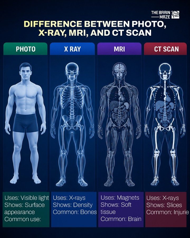
Medical Imaging: How Four Key Techniques Differ
Medical imaging plays a critical role in modern healthcare, allowing practitioners to peer inside the human body without invasive procedures. Four of the most commonly used techniques are photo, X-ray, MRI, and CT scans. Each of these technologies offers unique insights by utilizing different principles to visualize various aspects of the human anatomy.
Why This Matters
Understanding the differences between these medical imaging techniques is crucial for both healthcare professionals and patients. By knowing what each method reveals, patients can better understand their diagnoses and treatments. Healthcare professionals can choose the most appropriate imaging technique for a specific condition, ensuring accurate diagnoses and effective treatment plans.
Photo
Photos in a medical context are often taken to document the surface appearance of the body. This can include visible signs of injury, rashes, moles, or other skin conditions. Photos are a fundamental aspect of medical records and are often used to monitor changes in a patient's condition over time.
Uses
Photos use visible light to capture images. While this might seem simple, the surface appearance of the body can provide valuable information. Visible light is non-invasive and widely available, making it an accessible tool for medical professionals.
Common Uses
Skin examinations: Dermatologists and other medical professionals use photos to document skin conditions, track changes, and monitor the effectiveness of treatments.
Monitoring wounds and injuries: Photos help healthcare providers assess the healing process of wounds, burns, and other injuries.
Documentation: Photos are often used to document a patient's condition for medical records, providing a visual reference for future consultations.
X-Rays
X-rays have been a staple in medical imaging for over a century. They use a form of electromagnetic radiation to create images of the body. X-ray images, also known as radiographs, are particularly useful for visualizing bones and certain structures within the body.
Uses
X-rays are generated by a machine that produces a beam of X-ray photons, which pass through the body and are absorbed differently by various tissues. Bones, which are dense, absorb more X-rays than soft tissues, resulting in a contrast that creates a clear image.
Common Uses
Breast imaging: Mammograms are a type of X-ray used to detect breast cancer.
Chest imaging: Chest X-rays are used to diagnose conditions such as pneumonia, lung cancer, and tuberculosis.
Bone fractures: X-rays of broken bones help orthopedists plan for surgery or determine treatment.
MRI
Magnetic Resonance Imaging (MRI) is a powerful tool for imaging soft tissues, such as muscles, organs, and the brain. Unlike X-rays, MRI does not use ionizing radiation, making it a safer option for certain diagnostic needs.
Uses
MRIs use a combination of magnetic fields, radio waves, and computer technology to produce detailed images of the body. These images can be viewed in multiple planes, providing a comprehensive view of internal structures.
Common Uses
Brain imaging: MRIs are often used to diagnose conditions affecting the brain, such as tumors, aneurysms, and multiple sclerosis.
Musculoskeletal imaging: MRIs can show detailed images of muscles, ligaments, tendons, and cartilage, making them useful for diagnosing sports injuries or conditions like arthritis.
Organ imaging: MRIs are valuable for examining soft tissues and internal organs, such as the liver, kidneys, and intestines.
CT Scan
A Computed Tomography (CT) scan is a combination of X-rays and computer technology, providing detailed, cross-sectional images of the body. CT scans offer a more detailed view of the internal structures compared to traditional X-rays.
Uses
CT scans use multiple X-ray beams from different angles to produce a 3D image of the body. This technique is particularly useful for diagnosing complex conditions and planning surgeries.
Common Uses
Head and neck imaging: CT scans are used to diagnose conditions such as stroke, brain tumors, and sinus infections.
Chest and abdomen imaging: CT scans can diagnose conditions affecting the lungs, heart, liver, and other abdominal organs.
Bone and joint imaging: CT scans can provide detailed images of bones and joints, aiding in the diagnosis of fractures, arthritis, and other bone conditions.
Practical Tips
When patients undergo medical imaging, they may have questions or concerns about what to expect. These tips can help prepare for the process:
Preparation
- Hydration: For certain MRI scans, especially those involving contrast dyes, patients may need to stay hydrated. For CT scans, patients are encouraged to avoid liquids for a few hours before the exam, depending on the type of scan.
- Clothing: Wear comfortable, loose-fitting clothing without metal zippers, buckles, or snaps, as these can interfere with the imaging process.
- Jewelry and Accessories: Remove any jewelry, body piercings, or accessories, as metal objects can affect the quality of the images.
- Medications: Inform the healthcare provider about any medications or supplements being taken, as some may need to be adjusted before the scan.
During the Procedure
- Stay Still: Patients need to remain still during the procedure to ensure clear images. Movement can blur the images, leading to the need for additional scans.
- Communication: If the patient feels uncomfortable or experiences any discomfort, they should inform the healthcare provider immediately. Some patients may feel claustrophobic during MRI scans, so facilities offer open MRI machines or sedation if necessary.
- Follow Instructions: Healthcare providers will give specific instructions before and during the procedure. Following these instructions ensures the best possible results.
After the Procedure
- Hydration: After a CT scan or MRI with contrast, it is essential to stay hydrated. Drinking plenty of fluids helps the body eliminate the contrast agent.
- Rest: Patients may feel tired after the procedure, so taking it easy for the rest of the day is advisable.
- Follow-Up: Schedule any necessary follow-up appointments and communicate any concerns or questions to the healthcare provider.
Important Takeaways
Medical imaging techniques each offer unique insights into the human body, allowing healthcare professionals to diagnose and treat a wide range of conditions. Understanding the differences between these techniques can help patients make informed decisions about their healthcare and better understand their diagnoses.
Conclusion
The differences between photo, X-ray, MRI, and CT scan techniques are crucial for both healthcare providers and patients. By understanding what each method reveals, patients can better navigate their healthcare journey, and healthcare providers can choose the most appropriate imaging technique for accurate diagnoses and effective treatments. From visualizing surface conditions to diagnosing complex internal issues, each imaging method plays a vital role in modern healthcare, ensuring comprehensive and precise patient care.
Watch the Reel
Questions readers ask
What are the primary differences between an X-ray and an MRI scan?
X-rays use ionizing radiation to produce images of the body's internal structures, particularly bones, and are effective for detecting fractures or abnormalities. MRI scans, on the other hand, use magnetic fields and radio waves to create detailed images of soft tissues, making them ideal for examining organs, muscles, and ligaments.
How does a CT scan work and what makes it different from an MRI?
A CT scan combines a series of X-ray images taken from different angles to produce cross-sectional images of the body. Unlike MRI, which uses magnetic fields, CT scans are faster and often used for detecting internal injuries, tumors, or blood clots. However, they expose the patient to more radiation than a single X-ray.
What can I expect during a medical imaging photo session?
Medical imaging photos, often referred to as dermatoscopic images, capture the surface appearance of the skin. During a photo session, expect the healthcare professional to use a specialized camera to take close-up images of specific skin areas, often for monitoring moles or lesions.
Why might a doctor order an MRI brain scan?
An MRI brain scan is typically ordered to examine the brain's soft tissues in detail. This helps in diagnosing conditions such as tumors, aneurysms, or multiple sclerosis. MRI is particularly useful for brain imaging because it provides high-resolution images without the use of ionizing radiation.
Which medical imaging technique is best for examining soft tissues, and why?
MRI is the best technique for examining soft tissues. MRI scans use strong magnetic fields and radio waves to generate detailed images of soft tissues, such as muscles, ligaments, and organs, which are not clearly visible with other imaging methods like X-rays or CT scans.
What are the common medical imaging techniques used in healthcare?
The most common medical imaging techniques are photo, X-ray, MRI, and CT scans. Each technique is chosen based on the specific needs of the diagnosis, with photos capturing surface details, X-rays showing bone structures, MRI revealing soft tissues, and CT scans providing cross-sectional views of the body.
Recent articles
Fresh deep dives from the latest Reels we unpacked.



Comments
Be the first to comment.