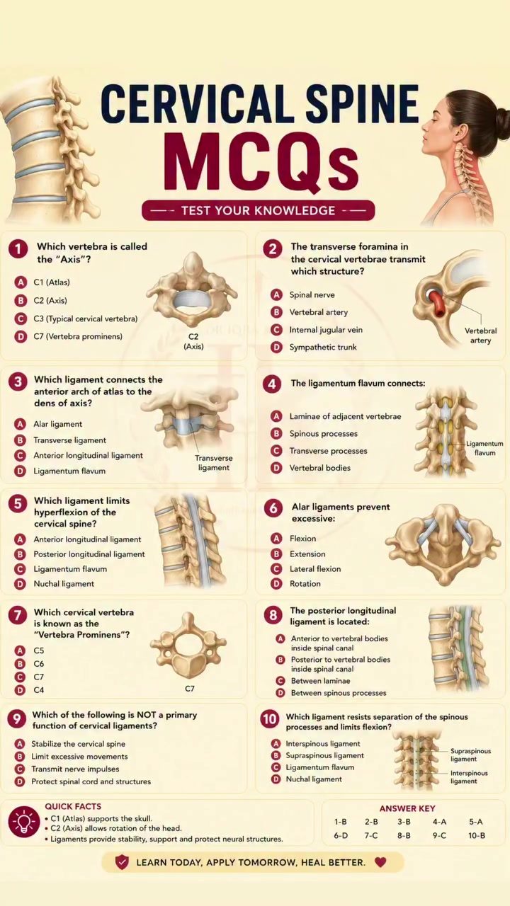
Cervical Spine Anatomy Quiz
Understanding the structure and function of the cervical spine is a cornerstone of medical and physiotherapy education. The cervical spine consists of seven vertebrae (C1-C7), each with unique features and roles. Here is an in-depth look based on the exam and key concepts covered in the quiz, which is designed to reinforce your knowledge of this critical area of the body.
Why this matters
The cervical spine is not just a stack of bones; it houses critical structures like the spinal cord and vertebral arteries. Moreover, its role in supporting the head and facilitating movement of the neck makes it a pivotal area of study for medical students, physiotherapists, and healthcare professionals. A solid understanding of cervical spine anatomy is essential for accurate diagnoses, effective treatments, and successful rehabilitation strategies. This quiz covers key aspects of cervical spine anatomy, including vertebral identification, ligament functions, and spinal stability, which are all crucial for clinical practice.
Key Concepts
Cervical Vertebrae Identification
The cervical spine comprises seven vertebrae, each with distinct characteristics:
- C1 (Atlas): The first cervical vertebra, known as the Atlas, supports the skull and allows for nodding movements.
- C2 (Axis): The second cervical vertebra, or Axis, facilitates rotational movements of the head.
- C3 to C6: These vertebrae are known as typical cervical vertebrae and share similar structural features.
- C7 (Vertebra Prominens): The seventh cervical vertebra is easily identifiable by its prominent spinous process and is a key landmark in physical examinations.
Ligaments and Spinal Stability
Ligaments play a crucial role in maintaining the stability and mobility of the cervical spine. Key ligaments include:
- Ligamentum Flavum: This connects the lamina of adjacent vertebrae, providing stability and preventing excessive flexion.
- Alar Ligaments: These prevent excessive flexion and rotation of the cervical spine, connecting the dens of the axis to the occipital condyles.
- Transverse Ligament: This connects the dens of the axis to the atlas, providing stability to the C1-C2 complex.
Vertebral Artery
The cervical vertebrae, particularly C1 to C6, have transverse foramina that transmit the vertebral artery. This artery supplies blood to the brainstem and cerebellum, making it a vital structure in the cervical spine.
Practical Tips
Effective Study Techniques
To reinforce your understanding of cervical spine anatomy, consider the following study techniques:
- Active Recall: Use flashcards or apps that require you to actively recall information rather than just recognize it.
- Practice Questions: Regularly practice multiple-choice questions (MCQs) to test your knowledge and identify areas that need improvement.
- Mnemonic Devices: Create mnemonic devices to remember the unique features of each cervical vertebra and the functions of key ligaments.
- Hands-On Learning: Use models or anatomical charts to visualize the structures and their relationships.
Clinical Application
- Physical Examination: Understanding the landmarks and structures of the cervical spine is essential for conducting accurate physical examinations and diagnosing conditions like cervical spondylosis, whiplash, or spinal cord compression.
- Rehabilitation: Knowledge of the cervical spine is crucial for designing effective rehabilitation programs, particularly for patients with neck injuries or spinal disorders.
Important Takeaways
- Structure and Function: The cervical spine's structure directly influences its function. Understanding this relationship is key to diagnosing and treating conditions.
- Clinical Relevance: The cervical spine is a clinically relevant area, and a thorough understanding of its anatomy is crucial for healthcare professionals.
- Practical Application: Regular practice with MCQs and hands-on learning can significantly enhance your understanding and retention of cervical spine anatomy.
Conclusion
Mastering cervical spine anatomy involves more than just memorizing facts; it requires a deep understanding of how structure relates to function and clinical conditions. Through active learning techniques, regular practice, and hands-on experience, you can strengthen your knowledge and apply it effectively in your clinical practice. Whether you're a medical student, physiotherapy student, or healthcare professional, a solid grasp of cervical spine anatomy is indispensable for providing the best possible care to your patients.
Watch the Reel
Recent articles
Fresh deep dives from the latest Reels we unpacked.




Comments
Be the first to comment.